UA-45667900-1

Monday, 14 March 2022

Fenamates (Diclofenac, Ponstan etc): certainly for Alzheimer’s, maybe some Epilepsy, but Autism? I’m Impressed!


Some readers of this blog are interested in the potential of mefenamic acid (MFA), sold as Ponstan, to treat autism. There is a lack of evidence currently. 

On the other hand, the evidence looks pretty overwhelming in the case of this class of drug to treat Alzheimer’s, hence today’s post. If you have a case of epilepsy at home, you can follow up on that loose end I left.

I also introduce MFA as a therapy for sound sensitivity and Misophonia. It was pretty impressive in the case of Monty, aged 18 with ASD.


The highlights are:


·        Fenamate NSAIDs reduce the incidence of Alzheimer’s

·        Fenamate NSAIDs delay the progression of those already with Alzheimer’s

·        Acetaminophen/Paracetamol worsens the progression of Alzheimer’s

·        Low dose aspirin is chemoprotective, as well as reducing blood clots that cause heart attack and stroke, but offers no Alzheimer’s benefit

·        MFA/Ponstan is very effective in reducing Monty’s sound sensitivity


The caveats 

As is always the case, there are caveats.

It is well known that low dose aspirin can cause dangerous bleeding events in specific sub-populations.

A study of 6 million people in Denmark showed that older people taking the Fenamate Diclofenac has a slightly higher risk of heart problems than other NSAIDs. The risk is actually very low and symptoms in those affected generally appear within a month (and disappear on cessation).


Incidence of Alzheimer’s


 Source:  https://www.intechopen.com/chapters/43129


The longer you live, the chance of developing Alzheimer’s rapidly increases. 

The signs are actual visible in a CT scan decades before the symptoms are evident.

Almost two-thirds of Americans with Alzheimer's are women.

Older Black Americans are about twice as likely to have Alzheimer's or other dementias as older Whites.

Of those with I/DD (Intellectual or Developmental Disability), it is people with Down Syndrome who are at major risk of early onset Alzheimer’s.  More than 50% will develop Alzheimer's. 


Non drug methods to protect against Alzheimer’s and other dementia 

In this blog we have encountered numerous dietary methods associated with reduced risk of all types of dementia and Alzheimer’s specifically.

·        Dietary nitrates (beetroot, spinach etc)

·        Betanin (the pigment in beetroot)

·        Ergothioneine (from mushrooms)

·        Spermidine (from wheatgerm and mushrooms)

·        Anthocyanin pigments from superfoods (bilberry, blueberry, purple sweet potato etc)


Maintaining normal blood pressure, blood glucose levels and cholesterol levels are big advantages. Normal body mass and regular exercise are also important.

Fenamates are a class of NSAID pain medication that many people have at home. In the US there are 10 million prescriptions a year of Diclofenac / Voltaren.

Another common Fenamate is Mefenamic Acid (MFA), commonly sold as Ponstan.  Ponstan is only expensive in North America. 

Most people’s reaction would be “Ah, yes those are pain medications, how could they help Alzheimer’s or other neurological conditions. Aren’t they the ones with those GI side effects?” 

NSAIDS deaden pain by blocking an enzyme called cyclooxygenase-2 (COX-2). Unfortunately, they also block to some extent a very similar enzyme called  cyclooxygenase-1 (COX-1). COX-1 promotes the production of the natural mucus lining that protects the inner stomach and contributes to reduced acid secretion.  Blocking COX-1 will cause GI side effects. Most people want to take an NSAID that is selective for COX-2.


Low dose Aspirin – the good COX-1 effect

There is a good effect from blocking COX-1, as from low dose aspirin (LDA), because it stops blood platelets sticking together and blocking blood flow.  LDA is also substantially chemoprotective and nobody has figured out why and it likely has nothing to do with COX1 or COX2. 

“Ishikawa et al. analyzed 51 randomized controlled trials (RCTs) and the cumulative evidence strongly supports the hypothesis that daily use of aspirin results in the prevention of cardiovascular disease (CVD), as well as a reduction in cancer-associated mortality [3].”


Anti-inflammatories in Alzheimer’s disease—potential therapy or spurious correlate? 

Epidemiological evidence suggests non-steroidal anti-inflammatory drugs reduce the risk of Alzheimer’s disease. However, clinical trials have found no evidence of non-steroidal anti-inflammatory drug efficacy. This incongruence may be due to the wrong non-steroidal anti-inflammatory drugs being tested in robust clinical trials or the epidemiological findings being caused by confounding factors. Therefore, this study used logistic regression and the innovative approach of negative binomial generalized linear mixed modelling to investigate both prevalence and cognitive decline, respectively, in the Alzheimer’s Disease Neuroimaging dataset for each commonly used non-steroidal anti-inflammatory drug and paracetamol. Use of most non-steroidal anti-inflammatories was associated with reduced Alzheimer’s disease prevalence yet no effect on cognitive decline was observed. Paracetamol had a similar effect on prevalence to these non-steroidal anti-inflammatory drugs suggesting this association is independent of the anti-inflammatory effects and that previous results may be due to spurious associations. Interestingly, diclofenac use was significantly associated with both reduce incidence and slower cognitive decline warranting further research into the potential therapeutic effects of diclofenac in Alzheimer’s disease.


Diclofenac Use Slows Cognitive Decline in Alzheimer Disease 

CHICAGO — While most common non-steroidal anti-inflammatory drugs (NSAIDs) do not significantly affect cognitive decline in patients with Alzheimer disease or mild cognitive impairment, research presented at the 2018 Alzheimer’s Association International Conference, held July 22-26, 2018, in Chicago, Illinois suggests that diclofenac actually reduces cognitive deterioration, while paracetamol accelerates decline. 

The study investigated cognitive decline associated with NSAID use in 1619 patients from the Alzheimer’s Disease Neuroimaging Initiative dataset. The Mini-Mental State Examination and the Alzheimer disease assessment scale were used to evaluate cognitive functioning. Additional variables that potentially explain cognitive decline were identified for the cohort including gender, apolipoprotein E genotype, level of education, vascular disorders, diabetes, and medication use. 


Study results showed that most common NSAIDs, including aspirin, ibuprofen, naproxen, and celecoxib did not alter cognitive degeneration in patients with mild cognitive impairment or Alzheimer disease. Diclofenac was the only NSAID that demonstrated a correlation with a slower rate of cognitive decline (ADAS χ2=4.0, P =.0455, MMSE χ2=4.8, P =.029). Conversely, paracetamol was correlated with accelerated cognitive deterioration (ADAS χ2=6.6, P =.010, MMSE χ2=8.4, P =.004), as well as apolipoprotein E ε4 genotype (ADAS χ2=316.0, P <.0001, MMSE χ2=191.0, P <.0001). 

Diclofenac’s correlation with slowed cognitive deterioration provides “exciting evidence for a potential disease modifying therapeutic,” the study authors wrote. If paracetamol’s deleterious effects are confirmed to be causative, it “would have massive ramifications for the recommended use of this prolific drug.”


One reason why paracetamol use might harm Alzheimer’s brains is the same reason it harms autistic brains; it depletes the level of the key antioxidant glutathione (GSH).  GSH will be in big demand in a damaged brain. 

As we will see later in this post, Fenamate class NSAIDs affect numerous ion channels, specifically Kv7.1, as a result some people with heart conditions will get side effects linked to arrhythmia and should therefore discontinue use.


Common painkiller linked to increased risk of major heart problems: Time to acknowledge potential health risk of diclofenac and reduce its use, say researchers -- ScienceDaily

Common painkiller linked to increased risk of major heart problems

Time to acknowledge potential health risk of diclofenac and reduce its use, say researchers 

The commonly used painkiller diclofenac is associated with an increased risk of major cardiovascular events, such as heart attack and stroke, compared with no use, paracetamol use, and use of other traditional painkillers, a new study finds.


The risk is actual quite low and is going to appear straight away, in terms of arrhythmia. If any drug or supplement makes you feel unwell, stop taking it and tell your doctor.


Which Fenamate for Alzheimer’s?

To decide which Fenamate is best for Alzheimer’s and indeed which might be helpful in some autism, it helps to ponder the various modes of action unrelated to COX-1 and COX-2. 

We have the NLRP3 inflammasome, which is suggested as the mechanism in Alzheimer’s.

Here we want to block inflammatory messenger like IL-1beta. In the chart below we see that Ibuprofen is useless, Diclofenac has an effect, Mefenamic acid is better, but Meclofenamic acid is the star.


Fenamate NSAIDs inhibit the NLRP3 inflammasome and protect against Alzheimer’s disease in rodent models

Non-steroidal anti-inflammatory drugs (NSAIDs) inhibit cyclooxygenase-1 (COX-1) and COX-2 enzymes. The NLRP3 inflammasome is a multi-protein complex responsible for the processing of the proinflammatory cytokine interleukin-1β and is implicated in many inflammatory diseases. Here we show that several clinically approved and widely used NSAIDs of the fenamate class are effective and selective inhibitors of the NLRP3 inflammasome via inhibition of the volume-regulated anion channel in macrophages, independently of COX enzymes. Flufenamic acid and mefenamic acid are efficacious in NLRP3-dependent rodent models of inflammation in air pouch and peritoneum. We also show therapeutic effects of fenamates using a model of amyloid beta induced memory loss and a transgenic mouse model of Alzheimer’s disease. These data suggest that fenamate NSAIDs could be repurposed as NLRP3 inflammasome inhibitors and Alzheimer’s disease therapeutics.


Fenamates and Ion Channels

https://scholarworks.wm.edu/cgi/viewcontent.cgi?article=2671&context=aspubs


A very broad range of ion channels are affected by Fenamates.

Researcher Knut Wittkowski focuses on the effect on potassium channels in his theory that Fenamates can treat autism and prevent non-verbal autism if given to toddlers.


Fenamates actually affect numerous ion channels.


·        Chloride channels

·        Non-selective cation channels

·        Potassium channels (Kv 7.1 , KCa 4.2, K2p 2.1, K2p 4.1, K2p 10.1)

·        Opens large conductance calcium-activated K+ channels (BKCa channels) 

https://www.researchgate.net/publication/348973521_Pharmacological_inhibition_of_BKCa_channels_induces_a_specific_social_deficit_in_adult_C57BL6J_mice

“Genetic variants in large conductance voltage and calcium sensitive potassium (BKCa) channels have associations with neurodevelopmental disorders such as autism spectrum disorder, fragile X syndrome, and intellectual disability… These findings support the relationship between BKCa channel impairment and social behavior. This demonstrates a need for future studies which further examine the contribution of BKCa channels to social behavior, particularly during critical periods of development.


·        Sodium channels

·        Blockage of acid-sensing ion channels (ASICs), which are implicated in numerous disorders and had their own post.

https://epiphanyasd.blogspot.com/2017/08/acid-sensing-ion-channels-asics-and.html


Fig. 2. Ion channels targeted by flufenamic acid. Flufenamic acid produces inhibition or activation of ion channels. Colored bars near ionic channel name correspond to the estimated EC50 for flufenamic effect. References are provided within the text.

 Having noted the above graphic, which actually applies to the closely related flufenamic acid, a logical question is to ask about the effect of Flufenamic acid on seizures. 

Flufenamic acid shows promise as an epilepsy drug


I am not looking for a seizure therapy, so I leave that loose end for someone who is.


Conclusion

The best initial defence against dementia is good diet and exercise. Sometimes that will not be enough, because the healthier you are, the longer you will live and so the threat from dementia increases. Some people have genes that predispose them to dementia.

Since most of us struggle to follow diets like those of ultra healthy people in Okinawa, or on a Greek island, it might be worthwhile adding beneficial functional foods (neutraceuticals) to your existing diet.

I drink a small amount of beetroot juice daily, which is not such a hard step to take. In addition to benefits to your heart and brain, another benefit has just been discovered; now it improves the oral microbiome :-


Research suggests changes in mouth bacteria after drinking beetroot juice may promote healthy ageing 

“Our findings suggest that adding nitrate-rich foods to the diet – in this case via beetroot juice – for just ten days can substantially alter the oral microbiome (mix of bacteria) for the better.”


Many older people take NSAIDs to treat painful conditions like arthritis, switching to a Fenamate NSAID would not be a difficult option and would give some protection from Alzheimer’s.

People already diagnosed with Alzheimer’s currently do not have any effective therapies. Drugs like memantine exist, but are not so effective.  If I was in that position, I would want to take a low dose of Mefenamic Acid, if that was unavailable, I would settle for Diclofenac.

Diclofenac (25mg to 100mg) is prescribed in much lower doses than Mefenamic Acid (250 to 500mg tablets). We see that the effect on the NLRP3 inflammasome is actually far greater from Mefenamic Acid than Diclofenac. If the Alzheimer’s effect is via inhibiting the NLRP3 inflammasome, then you might expect that only a fraction of a standard capsule of Mefenamic would be needed.  That would then really reduce any GI side effects via the unwanted effect on COX-1 or any chance of arrhythmia. 

The ketone BHB, like fenamate NSAIDs, inhibits the NLRP3 inflammasome.  Since in Alzheimer’s the brain loses the ability to transport enough glucose across the blood brain barrier, ketones can also be used as a supplementary fuel for the brain. In one of my old posts on BHB I remember the doctor treating her husband with early onset Alzheimer’s with large doses of ketones – with some success.


And Autism?

Is Knut right that the potassium channel modulation from Mefenamic Acid will benefit autism, or at least a sub-set of severe autism? We do not know.

Mefenamic Acid (MFA) has so many biologic effects, I very much doubt Alzheimer’s is the only neurological condition where it could be beneficial. 

I should add that MFA undoubtedly will have negative effects in some people, this is inevitable.


Stop the noise !!

We did have a problem recently with extreme sound sensitivity. Monty, aged 18 with ASD, has had increasing sound sensitivity (Misophonia) for a year, but the only real issue was with sounds at mealtimes.  Over a recent weekend the sensitivity increased so much he could not sleep and also drank unusually large amounts of water (this also connects to K+).
 

The next day at school he had a geography exam and he was completely dysfunctional. Monty’s assistant had prewarned the teacher and she agreed that he can sit the exam again next week.   

Fortunately, in the meantime the problem has been now been fixed (see below).

I was suggested to take to Monty to a Neurologist, but since there is no Dr Chez where we live, I did ignore that idea. In mainstream neurology sound sensitivity is just something you have got to learn to live with, perhaps with some Cognitive Behavioral Therapy (CBT) or just a pair of ear defenders, or those noise-cancelling headphones.

I did experiment years ago on the effect of an oral potassium supplement on reducing sound sensitivity, so I have long considered potassium ion channels a prime target.

Both hearing and the processing of the inputs is highly dependent on potassium channels, so I did return to MFA.  It has also been a topic in some recent email exchanges and I have long had some unopened packs of MFA at home.  The answer would be found in the kitchen cabinet and not in the neurology department

In bumetanide responders the Na-K-2Cl cotransporter (NKCC1) is over-expressed; it mediates the “coupled electroneutral movement of 1Na+, 1K+, and 2Cl ions across the plasma membrane of neurons”. This means that with each two chloride ions entering the neuron, come one sodium ion and one potassium ion.


Source: https://www.frontiersin.org/articles/10.3389/fncel.2019.00048/full


In summary, bumetanide responders have too much chloride in their neurons, the bubble on the left, above.


Knut’s theory was put to me recently as “MFA works on reducing neuron excitation by opening K+ channels, emptying the cell, which in return fills up with Cl- “.

If this is the case, MFA would do the opposite of Bumetanide.

I actually think MFA’s effect is much more complex.

The original idea of Knut was to prevent severe non-verbal autism developing in toddlers, by blocking the progression of the disease. MFA was essentially a medium-term treatment for toddlers, until the critical periods in brain development were past.  It was not a treatment for teenagers, by then the damage would have been done.

I think changing the baseline level of K+ inside neurons is going to have many effects.  Changing the baseline level of Cl- has a profound impact on cognition.

Unfortunately, everything is interrelated and so nothing is simple.

I did try MFA to eradicate the extreme sound sensitivity. I was concerned it might reduce cognition, by raising intracellular chloride and undo the bumetanide effect.

The extreme sound sensitivity did disappear following a day or two of starting 250mg a day of MFA, but that may just have been a coincidence.  The more mild sound sensitivity, that we had all learned to live with for months, also vanished; I do not see how that could be a coincidence.  Mood also became very good, perhaps a bit uncontrollably happy.

The next question is what happens to sound sensitivity when I stop giving MFA.  Time will tell, but so far the benefits have been maintained.

Sound sensitivity/Misophonia is a classic feature of autism;  TV depictions often portray a lonely looking boy wearing ear defenders. For many with Asperger’s misophonia is their main troubling issue. None of these people are taking bumetanide.  Monty has taken Bumetanide for nearly 10 years and never needed ear defenders.

You, like Prof Ben-Ari, might wonder if bumetanide use might cause a problem with potassium and hence hearing.  There is indeed a known risk of ototoxicity, which is actually a rare but possible side effect of loop-diuretic use, particularly furosemide.

Fluid in the inner ear is dependent upon a rich supply of potassium, especially in that part of the ear that translates the noises we hear into electrical impulses the brain interprets as sound.


Source: http://www.cochlea.eu/en/cochlea/cochlear-fluids


“Endolymph (in green) is limited to the scala media (= cochlear duct; 3), is very rich in potassium, secreted by the stria vascularis, and has a positive potential (+80mV) compared to perilymph.

Note that only the surface of the organ of Corti is bathed in endolymph (notably the stereocilia of the hair cells), whilst the main body of hair cells and support cells are bathed in perilymph.”


It is important to maintain a high level of potassium (K+) in the endolymph.

How the potassium gets there is a little bit complicated but it relies on:

·        The NKCC1 transporter

·        Potassium channel Kir4.1

·        Potassium channel KCNQ1 (Kv7.1) and in particular subunit KCNE1


Bumetanide blocks NKCC1 and so can potentially reduce potassium in the endolymph. Very high dose bumetanide would indeed risk ototoxicity.

We saw earlier in this post that Fenamates affect Kv7.1.

It is very poorly documented in the research, but Fenamates also affect Kir4.1.


The cochlea functions like a microphone. The auditory nerve then runs from the cochlea, hopefully bathed in potassium, to a station in the brainstem. From that station, neural impulses travel to the brain – specifically the temporal lobe, containing the primary auditory complex, where sound is attached meaning and we “hear”.


The auditory cortex is highlighted in pink and interacts with the other areas highlighted above


   Angular Gyrus   Supramarginal Gyrus   Broca's Area   Wernicke's Area 


By James.mcd.nz - self-made - reproduction of combined images Surfacegyri.JPG by Reid Offringa and Ventral-dorsal streams.svg by Selket, CC BY-SA 4.0, https://commons.wikimedia.org/w/index.php?curid=3226132


The peripheral auditory system links the microphone/cochlea to the brain. The Primary Auditory Neurons begin in the cochlea and terminate in the Brainstem (in the Cochlear Nuclei). In these neurons potassium channels play a key role.  These channels include KNa1.1 and KNa1.2, which are regulated by intracellular Na+ and Cl, are found in a variety of neurons.

We assume that intracellular Cl is disturbed in bumetanide responsive autism.

Everything has to function to ensure normal hearing and with normal perception attached to that hearing.  Problems can arise in the cochlea (microphone) or in any of the above areas in the brain involved in transmitting or processing those signals.


Fenamates for some Aspie’s with Misophonia?

Misophonia has been covered in previous posts and we saw that therapies do exist in the research.  I think that there are multiple causes of sound sensitivity and likely also for those with Misophonia.

Low dose roflumilast was one interesting therapy, that works for some people but not others.  It does nothing for Monty regarding misophonia/sensory gating.

I wonder if some sound-troubled Aspies will respond to low dose MFA?


The top shelf

In our case, the answer to good health is usually found in the kitchen, but sometimes tucked away out of reach, up high at the back of a shelf, gathering dust, next to my stockpile of NAC.

There will be a dedicated post on sound issues in autism, which will draw everything together to include information from earlier posts.


and, not to forget, 


Danke vielmals Knut !

(Thanks to Knut!)


Monday, 7 March 2022

SEECA 2022 - Autism Conference in Belgrade June 2-4


Our reader Tatjana has been busy organizing an autism conference in Belgrade, for doctors and parents. 

It is called the South-East European Conference on Autism (SEECA) and takes place June 2 - 4.

Researchers and clinicians from around the world are coming to speak, many of whom have been mentioned previously in this blog. Dr Frye of folate and mitochondrial disorders, Dr Lemmonier with Bumetanide, Dr Krigsman the gastroenterologist, Dr Adams with FMT, Dr Antonucci with an overview of the many interventions that he finds useful. Plus others I have not previously mentioned in my blog, who will talk about PANS/PANDAS, Suramin, Stem Cell Treatment and more.

Tatjana has been busy.

Click on the link below to read all about it.


SEECA Conference


You can attend the conference online, if you are not able to travel to Belgrade.


Tuesday, 22 February 2022

From a SWAN to Chopra-Amiel-Gordon Syndrome and the emergence of “-like” syndromes, CNTNAP2 etc


SWAN (Syndrome Without A Name)


 Today’s post is complicated, it is aimed at people who:

·        are interested in genetic testing for autism 

·        are affected by miss-expression of the genes:-

·        ANKRD17

·        TCF4

·        CNTNAP2

·        NRXN1


It is one of those posts that could go on forever; the more you dig, the more you uncover and you wonder why other people (salaried researchers) are not doing this.

Today’s post is mainly about a gene called ANKRD17, but it does highlight more general principles about those genetic testing results, some parents strive to obtain.  It does look at downstream effects of Pitt Hopkins Syndrome and “Pitt Hopkins-like Syndrome”, which likely merge into mainstream autism.

Many single gene autisms have already been identified, some have names and some are still SWANs (Syndromes Without A Name). Some syndromes have long been identified,  but their biological basis had not been identified.  From last year, loss of function of the gene ANKRD17 became Chopra-Amiel-Gordon Syndrome.  

Our reader Mary’s whole exome sequencing (WEG) from a few years ago, for her daughter, has now been flagged up as carrying a mutation leading to Chopra-Amiel-Gordon Syndrome.

In effect, the mutation in ANKRD17 went from be of no confirmed relevance to autism, to being causal, thanks to Dr Chopra and her pals.

This highlights the weakness in the interpretation of genetic testing.  Any benchmark list of autism genes is just a work in progress; your mutation may not yet be there.

Another gene recently queried by a reader was CNTNAP2, this turns out to a key DEG (differentially expressed gene) of a syndrome with its own name, Pitt Hopkins Syndrome, caused by reduced expression of TCF4 (Transcription Factor 4).  Reduced expression of TCF4 has very many effects, but one effect is to reduce expression of CNTNAP2.

In lay-speak, lack of TCF4 causes a cascade of effects, one of which is on the expression of CNTAP2.  We see that people with a CNTAP2 mutation share many of the features of people having a TCF4 mutation.  So, of all the many effects caused by TCF4, those along the TCF4-CNTAP2 pathway should be focused on.  The mutation in CNTNAP2, quite rationally, is now called Pitt Hopkin-like Syndrome-1.  There is also a Pitt Hopkin-like Syndrome-2 which is caused by a mutation in NRXN1 (neurexin 1). 


https://royalsocietypublishing.org/doi/pdf/10.1098/rsob.210091


In mammals, the neurexins are encoded by three NRXN genes (NRXN1-3), each of which has both an upstream promoter that is used to generate the α-neurexins, and a downstream promoter that is used to generate the shorter β-neurexins [13,15].


α-neurexins are composed of six large extracellular laminin/neurexin/sex hormone-binding (LNS) globulin domains with three interspersed epidermal growth factor (EGF)-like regions


Just note the term EGF.


In very recent research we see that a reduction in epithelial growth factor may be what is driving some of the key clinical features, such as lack of language.


Role of CNTNAP2 in autism manifestation outlines the regulation of signaling between neurons at the synapse

CNTNAP2 has been identified as a master gene in autism manifestation responsible for speech-language delay by impairing the EGF protein domain and downstream cascade. The decrease in EGF is correlated with vital autism symptoms, especially language disabilities.

Autism exhibits genetic heterogeneity, and hence, it becomes difficult to pinpoint one single gene for its manifestation. The gene clusters with varied pathways show the convergence of multiple gene variants, resulting in autism manifestation. Whole-exome sequencing proves to be a reliable tool for deciphering the causal genes for autism manifestation. Deciphering the autism exome identified the mutational landscape derived from single and multi-base DNA variants. Genes carrying mutations were identified in synaptogenesis processes, EGF signaling, and PI3K/MAPK signaling. Protein-protein interactions of NrCAM and CNTN4 with CNTNAP2 increased the impact and burden on autism.


Shining a light on CNTNAP2: complex functions to complex disorders

TCF4 encodes a basic helix-loop-helix (bHLH) transcription factor that binds near the start site of CNTNAP2 to upregulate its expression (Figure 1a).48 In humans, TCF4 is more highly expressed in the neocortex and hippocampus than in the striatum, thalamus and cerebellum.49 Mutations in TCF4 have been shown to cause Pitt–Hopkins syndrome (PTHS) and three rare TCF4 SNPs are associated with schizophrenia.17495051 PTHS is characterised by severe intellectual disability, absent or severely impaired speech, characteristic facial features and epilepsy.52 Many of these features are shared with patients carrying CNTNAP2 mutations, leading researchers to test patients with PTHS-like features for CNTNAP2 mutations.17 Two mutations affecting the CNTNAP2 locus (one homozygous and one compound heterozygote) were identified in two independent pedigrees (Table 1). This suggested that disruption of the TCF4–CNTNAP2 pathway could be related to intellectual disability, seizures, and/or behavioural abnormalities.


One of our readers in Australia recently queried the potential significance of a mutation (an SNP) in CNTNAP2.  Based on the above, it clearly could be very important. 

What is the common link between TCF4, CNTNAP2 and NRXN1? It would seem to be EGF (epidermal growth factor).

It looks quite possible that EGF is disturbed in much broader autism. It appears that inflammation may reduce EGF levels. It is a rather circular argument, but we also know that EGF reduces inflammation.

To sum up people, with autism likely want more EGF and we already knew that they definitely want less inflammation.  

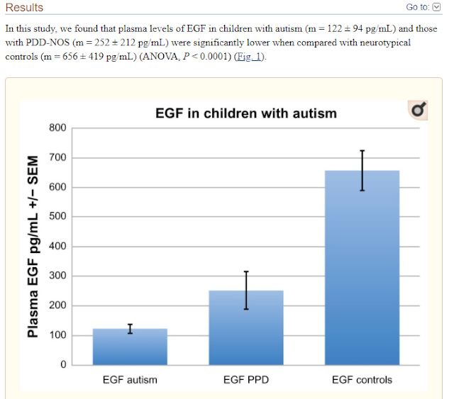
Decreased Epidermal Growth Factor (EGF) Associated with HMGB1 and Increased Hyperactivity in Children with Autism

These results suggest an association between decreased plasma EGF levels and selected symptom severity. We also found a strong correlation between plasma EGF and HMGB1, suggesting inflammation is associated with decreased EGF.


ANKRD17 

Finally, we get back to ANKRD17. 

Our reader Mary has already highlighted this recent paper: - 

Heterozygous ANKRD17 loss-of-function variants cause a syndrome with intellectual disability, speech delay, and dysmorphism


Dysmorphic facial features of the ANKRD17-related disorder


ANKRD17 is an ankyrin repeat-containing protein thought to play a role in cell cycle progression, whose ortholog in Drosophila functions in the Hippo pathway as a co-factor of Yorkie. Here, we delineate a neurodevelopmental disorder caused by de novo heterozygous ANKRD17 variants. The mutational spectrum of this cohort of 34 individuals from 32 families is highly suggestive of haploinsufficiency as the underlying mechanism of disease, with 21 truncating or essential splice site variants, 9 missense variants, 1 in-frame insertion-deletion, and 1 microdeletion (1.16 Mb). Consequently, our data indicate that loss of ANKRD17 is likely the main cause of phenotypes previously associated with large multi-gene chromosomal aberrations of the 4q13.3 region. Protein modeling suggests that most of the missense variants disrupt the stability of the ankyrin repeats through alteration of core structural residues. The major phenotypic characteristic of our cohort is a variable degree of developmental delay/intellectual disability, particularly affecting speech, while additional features include growth failure, feeding difficulties, non-specific MRI abnormalities, epilepsy and/or abnormal EEG, predisposition to recurrent infections (mostly bacterial), ophthalmological abnormalities, gait/balance disturbance, and joint hypermobility. Moreover, many individuals shared similar dysmorphic facial features. Analysis of single-cell RNA-seq data from the developing human telencephalon indicated ANKRD17 expression at multiple stages of neurogenesis, adding further evidence to the assertion that damaging ANKRD17 variants cause a neurodevelopmental disorder.


Neonatal growth parameters were normal in the majority of individuals (Table S2) but postnatal growth failure was a feature of almost half of the individuals (height < 2 SD in n ¼ 12 and weight < 2 SD in n ¼ 9). One individual with marked growth failure (individual 3, height 3.8 SD) was under treatment with growth hormone (GH), although GH stimulation testing was normal. Feeding difficulties, especially reduced oral intake, were reported at some stage in 11 individuals, 5 of whom required G-tube nutritional supplementation. Postnatal microcephaly (OFC < 2SD) was noted in seven individuals, and macrocephaly in four (one of these individuals, however, also harbored a pathogenic de novo NSD1 variant (GenBank: NM_022455.4, c.2615T>G [p.Leu872*]). Epilepsy was reported in nine individuals (individuals 1, 2, 16, 19, 21, 25, 27, 28, and 33), with an age of onset of under 2 years for five individuals (individuals 1, 2, 16, 19, and 25). Focal seizures with secondary generalization was the most common seizure subtype, present in five individuals (individuals 1, 2, 21, 25, and 27). One individual had Lennox-Gastaut epilepsy (individual 16), one had tonic seizures with head deviation (individual 19), one had mixed myoclonic and tonic-clonic epilepsy (individual 33), and another a mixture of tonic-clonic and absence seizures (individual 28). Seizures were well controlled (less frequent than every 2 years) in five individuals (individuals 2, 21, 25, 28, and 33), all of whom were on three or fewer antiepileptic drugs (AEDs). Moderate control, with seizures every 2–3 months, was reported in individual 1, who was on Valproate monotherapy. Two individuals had refractory epilepsy during at least parts of their disease course—individual 19 who had frequent tonic seizures in infancy that resolved with topiramate monotherapy and individual 16 Table 2. Frequencies of phenotypic characteristics of individuals with ANKRD17 variants Frequency Sex F ¼ 19, M ¼ 15 Growth Height < 2 SD 12/31 Weight < 2 SD 9/30 OFC < 2 SD 7/31 OFC > 2 SD 4/31 Development DD or ID 31/34 severe 7 moderate 12 mild 5 borderline 7 Motor delay 20/29 Speech delaya 29/32 Other ASD, n ¼ 8; ADHD, n ¼ 4 Neurology Epilepsy 9/33 Abnormal EEG 10/23 Brain MRI abnormalities 11/23 Gait or balance abnormalities 9/25 Spasticity or hypertonia 4/26 Other Recurrent infections 11/33 Feeding problems 11/27 Palate abnormalities 3/34 Hypermobility 9/29 Ophthalmological abnormalities 13/23 Miscellaneous Minor digital anomalies 6 Genitourinary abnormalities 5 Pigmentary abnormalities 4 Scoliosis 3 Abnormal bone mineralization 2 Prominent blood vessels 2 ADHD, attention deficit hyperactivity disorder; ASD, autism spectrum disorder a For details see Table S1 The American Journal of Human Genetics 108, 1138–1150, June 3, 2021 1143 who had multiple seizures every day despite three AEDs. Further details of epilepsy phenotype, including previously trialled AEDs, are noted in Table S2. There were four individuals without epilepsy in whom an abnormal EEG was recorded. Other neurological features include poor balance and/or abnormal gait (9/25) and peripheral spasticity (4/26, one of whom one was microcephalic). Neuroimaging abnormalities were identified in 11 of the 23 individuals in whom an MRI was recorded. Abnormalities include decreased white matter volume (individuals 14, 16, and 18), thinning of the corpus callosum (individuals 14 and 19), optic nerve hypoplasia (individuals 18 and 19), a localized hyperintensity (individuals 7 and 31), right temporal sclerosis (individual 16), dilated Virchow-Robin spaces (individual 6), periventricular nodular heterotopia (individual 30), and an arachnoid (individual 24) and pineal cyst (individual 16). Ophthalmological abnormalities were reported in 13/23 individuals. There were nine individuals with recurrent bacterial infections, one with recurrent viral infections, and one individual with recurrent infections that were both viral and bacterial. The source of bacterial infection was primarily the upper and lower respiratory system and the middle ear (nine individuals) and in some cases required hospitalization. Two individuals were on low-dose prophylactic antibiotics for recurrent otitis media or respiratory tract infections. Notably, individual 26 had a history of pseudomonas and methicillin-resistant staphylococcal aureus (MRSA) infection on his toes. Immunology assessments were recorded in five individuals, details of which can be found in Table S2, with no obvious immunodeficiency identified in these individuals. Generalized joint hypermobility was reported in 9/29 individuals. Notably, there were two individuals with cleft palate in the context of Pierre Robin sequence (PRS) and another with cleft lip and palate. Other infrequent features include minor digital anomalies (n ¼ 6), genitourinary abnormalities (n ¼ 5, of whom three had unilateral renal agenesis), abnormal skin pigmentation (n ¼ 4), scoliosis (n ¼ 3), abnormality of bone mineralization (n ¼ 2), and cutaneous prominence of blood vessels (n ¼ 2). Figure 2 shows the facial features of individuals with the ANKRD17-related neurodevelopmental disorder. Key dysmorphic features include a triangular-shaped face found in 10 of the 24 individuals for whom photos were available with a high anterior hairline (19/24), eyes which are either deep-set (5/24) or almond shaped (8/24) with periorbital fullness (6/24), thick nasal alae and flared nostrils (9/24), full cheeks (7/24), and a thin upper lip (12/24). The degree of dysmorphism was variable, with several individuals (particularly individuals 8 and 10) presenting with only subtle dysmorphic characteristics. Persistence of the high anterior hairline, periorbital fullness, and full cheeks into adulthood is demonstrated in individual 12 (age 30 years) and individual 25 (age 34 years). A number of diagnoses had been considered in several individuals prior to the identification of an ANKRD17 variant, including SATB2-associated syndrome (MIM: 612313) in individual 5 who presented with PRS, triangular facies and speech delay, and Floating-Harbour syndrome (MIM: 136140) in individual 9 who presented with marked short stature (height < 3 SD), microcephaly (head circumference < 2.5 SD), dysmorphic features, and borderline ID. This highlights the phenotypic overlap of the ANKRD17-related disorder with a number of other genetic syndromes, notably those with expressive language delay. In our cohort, significant speech delay was reported in most individuals (n ¼ 29) even in those with IQ in the borderline range. The finding that verbal IQ was reduced relative to performance IQ in three of the five individuals for whom deep neuropsychological phenotyping was available adds further evidence to our observation that expressive language is particularly affected in this disorder


How were the 34 individuals identified?

In the Table 1 of the paper, we see that the great majority of the children had been identified from WES (whole exome sequencing), a few had WGS (whole genome sequencing) and just one via micro array testing.

They families clearly opted to share their data, in the hope of some researcher finding it useful later, as Chopra, Amiel and Gordon clearly did after a few years later.


How do you figure out the DEGs (differentially expressed genes)?

To treat ANKRD17 deficiency (now known as Chopra Amiel Gordon Syndrome) you have a choice.

·        Increase expression of ANKRD17 via gene therapy, or a drug (if that were possible)

·        Treat some of the downstream DEGs (Differentially Expressed Genes)

Mary asked how you could identify the DEGs, given there is only one paper published on Chopra Amiel Gordon Syndrome.

You can start by reviewing everything known about ANKRD17.

A very good place to start is on the GeneCards website.

https://www.genecards.org/cgi-bin/carddisp.pl?gene=ANKRD17


Most people will end up having to learn some new words to understand everything on the above website. 

The first thing to note is just how wide ranging are the functions of this gene and this accounts from the wide-ranging problems associated with it.  It even plays a role in dealing with both viral and bacterial infections.

It is particularly upregulated in the fetal brain and that likely leads to the autism/ID related effects.

Protein differential expression in normal tissues from HIPED for ANKRD17 Gene 

This gene is overexpressed in Lung (18.9), Platelet (15.4), Retina (8.4), and Fetal Brain (6.4).


We can see that this gene is associated with Chopra Amiel Gordon Syndrome and Non-Specific Syndromic Intellectual Disability.

Quite possibly, Non-Specific Syndromic Intellectual Disability was used as a term because Chopra Amiel Gordon Syndrome did not yet exist.

But is useful to look up Non-Specific Syndromic Intellectual Disability, to see which other genes are listed.  This then tells you much about what can cause ID.  Follow the link below. 

https://www.malacards.org/card/non_specific_syndromic_intellectual_disability

We see a very long list of syndromes and genes.

There are 61 genes listed.

Going back to the Genecards ANKRD17 page, we can see if there are known protein interactions that might result in autism/ID.


 For even more related genes/proteins you can look here

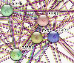
 https://string-db.org/cgi/network?taskId=b8Tr5wHDZSsf&sessionId=b1SfhFTkjWxd


EIF4E2 does look familiar, and I recall a link to Fragile X.  So, I looked it up.

Note that we see both EIF4E and EIF4E2 - Eukaryotic Translation Initiation Factor 4E Family Member 2.  Note that is has a second name, 4EHP. 

EIF4E2 is a version/homolog of EIF4E 

EIF4E2 = 4EHP 


The eIF4E homolog 4EHP (eIF4E2) regulates hippocampal long-term depression and impacts social behavior 

Background: The regulation of protein synthesis is a critical step in gene expression, and its dysfunction is implicated in autism spectrum disorder (ASD). The eIF4E homologous protein (4EHP, also termed eIF4E2) binds to the mRNA 5' cap to repress translation. The stability of 4EHP is maintained through physical interaction with GRB10 interacting GYF protein 2 (GIGYF2). Gene-disruptive mutations in GIGYF2 are linked to ASD, but causality is lacking. We hypothesized that GIGYF2 mutations cause ASD by disrupting 4EHP function.


4EHP is expressed in excitatory neurons and synaptosomes, and its amount increases during development. 4EHP-eKO mice display exaggerated mGluR-LTD, a phenotype frequently observed in mouse models of ASD. 


Conclusions: Together these results demonstrate an important role of 4EHP in regulating hippocampal plasticity and ASD-associated social behaviors, consistent with the link between mutations in GIGYF2 and ASD.


The disruption of protein synthesis (mRNA translation or translation) in the brain by genetic perturbations of its regulators constitutes a known underlying etiology for ASD [23]. For most mRNAs, initiation of translation requires binding of initiation factors to their 5′ end at a modified guanine nucleotide (m7GpppN, where N is any nucleotide) termed the 5′ cap [4]. The eukaryotic initiation factor (eIF) 4F complex is comprised of the cap binding protein eIF4E, an mRNA helicase eIF4A, and a molecular scaffold eIF4G. Together these proteins facilitate recruitment of the ribosomal 43S preinitiation complex to the mRNA. Overactivity of eIF4E in humans has been implicated in ASD [56] and ASD-like phenotypes in mice [78]. Indeed, disruption of the proteins regulating eIF4E activity, such as fragile X mental retardation protein (FMRP) [9], cytoplasmic FMR1 interacting protein 1 (CYFIP1) [10], and eIF4E-binding protein 2 (4E-BP2) [81112], is implicated in ASD. It is therefore necessary to investigate the function of ASD-linked genes that encode for regulators of translation. Whole-genome sequencing of ASD patients has been invaluable in identifying these genes.


If you look up the protein interaction for the Fragile X gene (FMRI), you do indeed see EIF4E close by.  FMR1 encodes the fragile X mental retardation protein.


This blog is full of ideas regarding treating Fragile X, because there are so many studies of that type of autism.

It is rather mind-boggling that there are still no approved therapies for Fragile X.  The same holds true for Down Syndrome (DS).  This is a recurring story, where it pays to be the early adopter, not one of the passive followers.


Leaky ATP from either Mitochondria or Neurons in Fragile X and Autism

l

In that post I suggested Mirapex - a miracle for Fragile X?”


In the post below we saw how EIF4E leads to autism, and how FMRP from Fragile-X affects EIF4E.


Vasopressin, Oxytocin, the Lateral Septum, Aggression and Social Bonding, Autism gene NLGN3 and MNK inhibitors for reversing Fragile-X and likely more Autism


One of the papers below goes further and suggests

“This work uncovers an unexpected convergence between the genetic autism risk factor Nlgn3, translational regulation, oxytocinergic signalling, and social novelty responses”

“We propose that pharmacological inhibition of MNKs may provide a new therapeutic strategy for neurodevelopmental conditions with altered translation homeostasis”

“Our work not only highlights a new class of highly-specific, brain-penetrant MNK inhibitors but also expands their application from fragile X syndrome to a non-syndromic model of ASD”


Regarding Fragile X 

“Collectively, this work establishes eFT508 (an MNK inhibitor) as a potential means to reverse deficits associated with FXS.”


Conclusion 

My quick look at the subject suggests that, amongst other likely DEGs, the NLGN (neuroligin) genes are quite possibly miss-expressed.

In humans, alterations in genes encoding neuroligins are implicated in autism and other cognitive disorders.

In Genecards the association is with EIF4E2 rather than the EIF4E, which we know affects neuroligin expression. But EIF4E2 is just a version of EIF4E.

These protein interaction maps are not perfect and different sources often come up with slightly different maps.


What are Neurexins and Neuroligins?

Neurexins and neuroligins are synaptic cell-adhesion molecules that connect pre- and postsynaptic neurons at synapses, they mediate signalling across the synapse, and shape the properties of neural networks by specifying synaptic functions. Neurexins and neuroligins are therefore very important and can be dysfunctional in autism.

It looks like growth signaling is disturbed in Chopra-Amiel-Gordon Syndrome, but it not always in the same way. Both too much and too little growth are possible.

An MRI would not be a bad idea, and measuring the corpus callosum would be helpful. The corpus callosum connects the right and left side of the brain and is the largest white matter structure in the brain, which means lots of myelin should be there.

If it is very narrow, that would tell you something, hopefully it is normal.  You cannot really change its size, but if it lacked myelination that might be something you could affect.   

Positive Correlations between Corpus Callosum Thickness and Intelligence


Trying the cheap and partially effective treatments for fragile X might be helpful.  It is possible that the Fragile X DEGs overlap with the Chopra-Amiel-Gordon Syndrome DEGs.

The following drugs are cheap generics that are helpful, to some extent, in Fragile-X.

·        Metformin

·        Lovastatin

·        Baclofen


As the altered E/I balance is present in Fragile X and most autism, it would be worthwhile trying the E/I corrective therapies that exist, in case one is beneficial.  There are different causes of an E/I imbalance, but since there are not many therapies, it is easier to just try them one by one. 

It is also highly likely that common features of autism may be present, such as

·        oxidative stress (NAC)

·        neuroinflammation (numerous therapies)

·        impaired myelination (Clemastine, Ibudilast, NAG) NAG is not the same as NAC, it is N-acetylglucosamine

·        mitochondrial dysfunction (Carnitine, antioxidants, activate PGC-1 alpha via PPAR gamma e.g. with Pioglitazone)

·        folate receptor antibodies (Calcium folinate)


If the Corpus Callosum is smaller than it should be, or is demyelinated, you could try high bio-availability curcumin, in addition to the above pro-myelination therapies.

Which ion channel dysfunctions appear in Chopra-Amiel-Gordon Syndrome?  I did not see any clues, but where there is epilepsy, there is very likely going to be an ion channel dysfunction involved.